
Dog skeleton 101 Dog Anatomy Bones Animal Hackers
2021 Ultimate Guide to Dog Anatomy. As the pace of veterinary advancement accelerates, even the most experienced veterinary teams are challenged to keep up with all the changes that impact their practice.. potential outcomes and prognoses and tools e.g. anatomical diagrams, clinical formulas and programs and home care videos; Enhancing the.

Dog Skeleton Drawing at GetDrawings Free download
Anatomy of a Dog Dog anatomy details the various structures of canines (e.g. muscle, organ and skeletal anatomy). The detailing of these structures changes based on dog breed due to the huge variation of size in dog breeds. Would you be surprised to know that short dogs are more aggressive? Or taller dogs are more affectionate?

FileDog anatomy lateral skeleton view.jpg
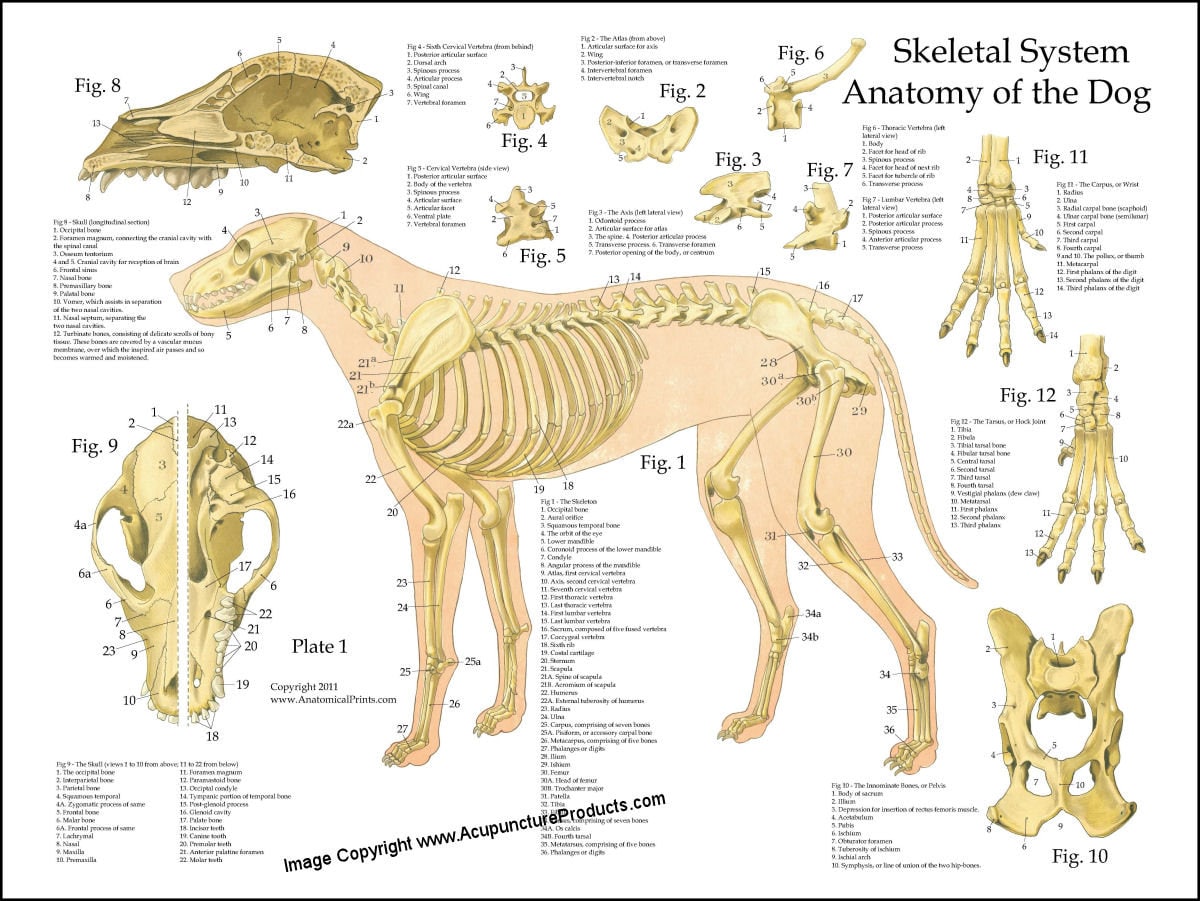
Labeled atlas of anatomy: illustrations of the dog: Bones - Skeletal system Dog - Muscles Dog - Thorax/Abdomen/Pelvis Animal - Anatomy atlas: Cardiovascular system Veterinary anatomy - Animal: ANATOMICAL PARTS Abdomen Abdominal aorta Abdominal mammary gland Abdominal mammary region Accessory carpal bone Acromion Adductor muscle

Dog Skeleton Anatomy by TheDragonofDoom on DeviantArt
Forelimb Hindlimb Joints Bone types and parts of the dog skeleton Regarding bone types, the dog skeleton is made of three main types of bones: long, irregular (no particular shape) and flat bones In the big picture, the dog skeleton is made of two basic parts: axial and appendicular (limbs).

Dog Anatomy Dog Skelton
Animal Anatomy (Veterinary Diagrams) Dog Skeletal Anatomy. High Resolution PDF for Printing. Click Here. Link to More Information About This Animal. Click Here. Citing Research References. When you research information you must cite the reference. Citing for websites is different from citing from books, magazines and periodicals.

Dog skeleton with major bone elements labeled (Davis, 1987, p. 54;... Download Scientific Diagram
Dog anatomy comprises the anatomical studies of the visible parts of the body of a domestic dog.Details of structures vary tremendously from breed to breed, more than in any other animal species, wild or domesticated, as dogs are highly variable in height and weight. The smallest known adult dog was a Yorkshire Terrier that stood only 6.3 cm (2.5 in) at the shoulder, 9.5 cm (3.7 in) in length.
Dog Skeletal Skull Anatomy Poster 18 X 24 Etsy
Dog Health 9 Dog Anatomy Illustrations With Facts & Additional Resources Brush up on your male and female dog anatomy with these beautiful, anatomical illustrations. By Kelly Roper Updated May 31, 2023 filadendron / E+ via Getty Images

Dog skeleton with major bone elements labeled (Davis, 1987, p. 54;... Download Scientific Diagram
This module of vet-Anatomy presents an atlas of the anatomy of the head of the dog on a CT. Images are available in 3 different planes (transverse, sagittal and dorsal), with two kind of contrast (bone and soft tissues).

Vintage 1935 Dog Veterinary Print Skeleton Of Dog Anatomy Of Dog Canine Skeleton Dog Bones Book
Diaphragm: The diaphragm is the primary muscle involved in breathing. When a dog barks, it contracts the diaphragm forcefully to expel air out of its lungs and through its vocal cords. Laryngeal muscles: The laryngeal muscles control the opening and closing of the dog's vocal cords, which are located in the larynx (voice box) in the neck.

Labeled atlas of anatomy illustrations of the dog Bones Skeletal system Dog skeleton
ISSN 2534-5087. This veterinary anatomy module of the dog contains 218 illustrations dedicated to the canine osteology anatomy. Here are presented scientific illustrations of the canine skeleton, with the main dog's bones and its structures displayed from different anatomical standard views (cranial, caudal, lateral, medial, dorsal, palmar..).

Dog skeleton Diagram Quizlet
A Visual Guide to Understanding Dog Anatomy With Labeled Diagrams Dog anatomy is not very difficult to understand if a labeled diagram is present to provide a graphic illustration of the same. That is exactly what you will find in this DogAppy article.

Anatomy of a male dog crosssection, showing the skeleton and internal organs. Colour process
Dog tongue anatomy with special features and labeled diagrams. Dog teeth anatomy. The teeth of a dog are the highly specialized structure in its mouth. You will find forty-two teeth in the mouth cavity of a dog. It is very important to know the eruption time of the dog's permanent teeth a veterinarian.
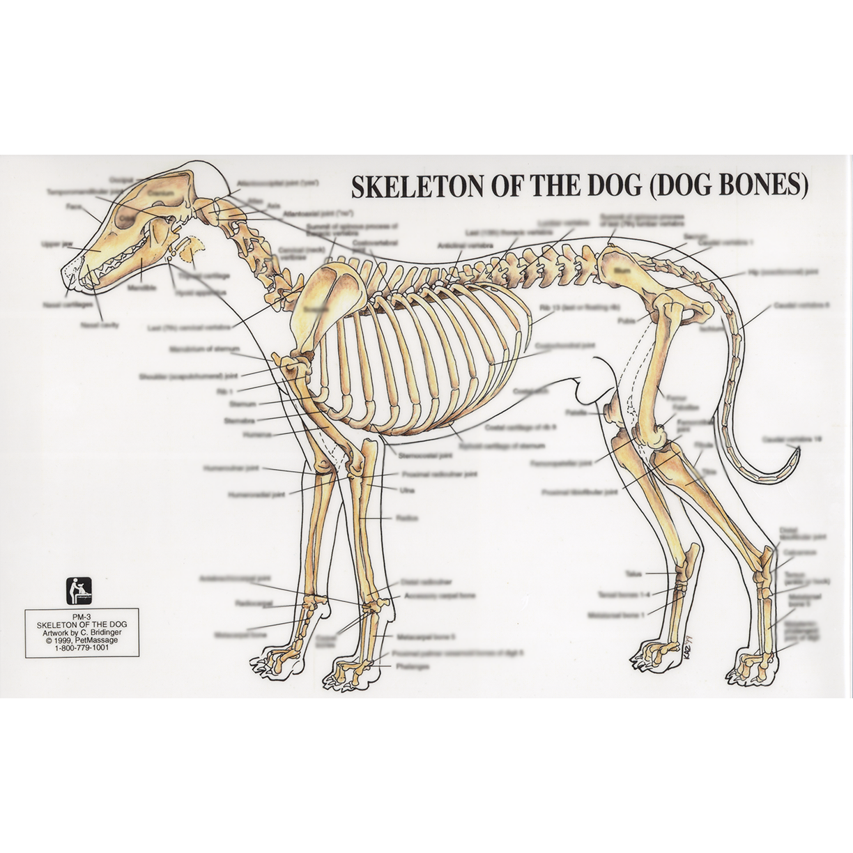
PetMassage™ Chart 3 Skeleton of the Dog · PetMassage™ Training and Research Institute
The Anatomage Dog is the first highly detailed dog anatomy atlas that comprehensively features internal organs, including vascular systems and muscular-skeletal structures. Originating from real dog data, the Anatomage Dog exhibits the highest level of anatomical accuracy. All of its volumetric 3D and individual structures are segmented, users.

Anatomy of dog skeleton with labeled inner bone scheme vector illustration Dog skeleton
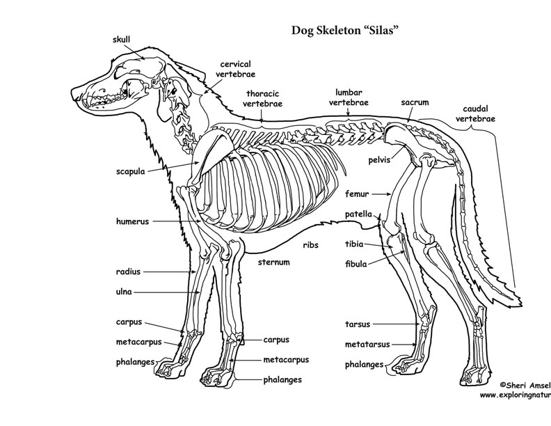
Dog Skeleton Anatomy with Labeled Diagram 25/04/2023 31/12/2021 by Sonnet Poddar The dog skeleton anatomy consists of bones, cartilages, and ligaments. You will find two different parts of the dog skeleton - axial and appendicular. Here, I will show you all the bones from the axial and appendicular skeleton with their special osteological features.
Dog Skeletal Anatomy
Here, I will provide a dog skeleton labeled diagram and the different parts of a dog diagram. In the dog skeleton labeled diagram, I tried to show you all the bones from the body. This might help you understand the different regions of the body so quickly. I would like to show different external features of a dog again here in a labeled picture.
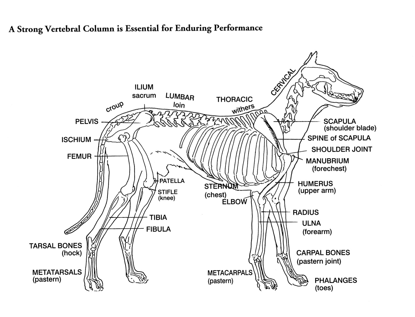
Helen King on Structure Evaluation Susan Garrett's Dog Training Blog
Dog skeleton. As with any vertebrate animal, the skeleton of a dog has the function of supporting the body for movement and protecting its internal organs. We can divide the canine skeleton into three main sections: Axial skeleton: skull, spine, ribs and sternum bones. Appendicular skeleton: bones of the extremities.